What research do we do?
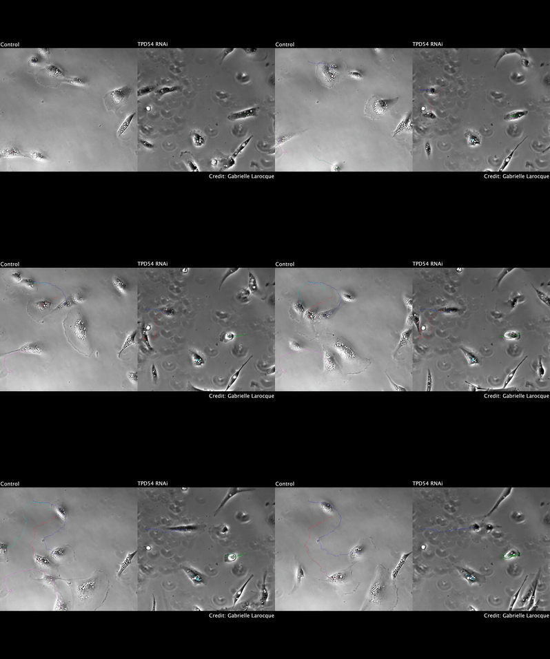
We are working on two things that cells do: how they divide and how they move stuff around inside themselves. These jobs are important and so any problems with them can lead to disease. This is why we are trying to understand these jobs, so that we can think of new ways to treat human diseases such as cancer or heart disease.
We publish articles in scientific journals about our research. These can be quite technical, so we write accessible summaries of each one here.
Membrane trafficking
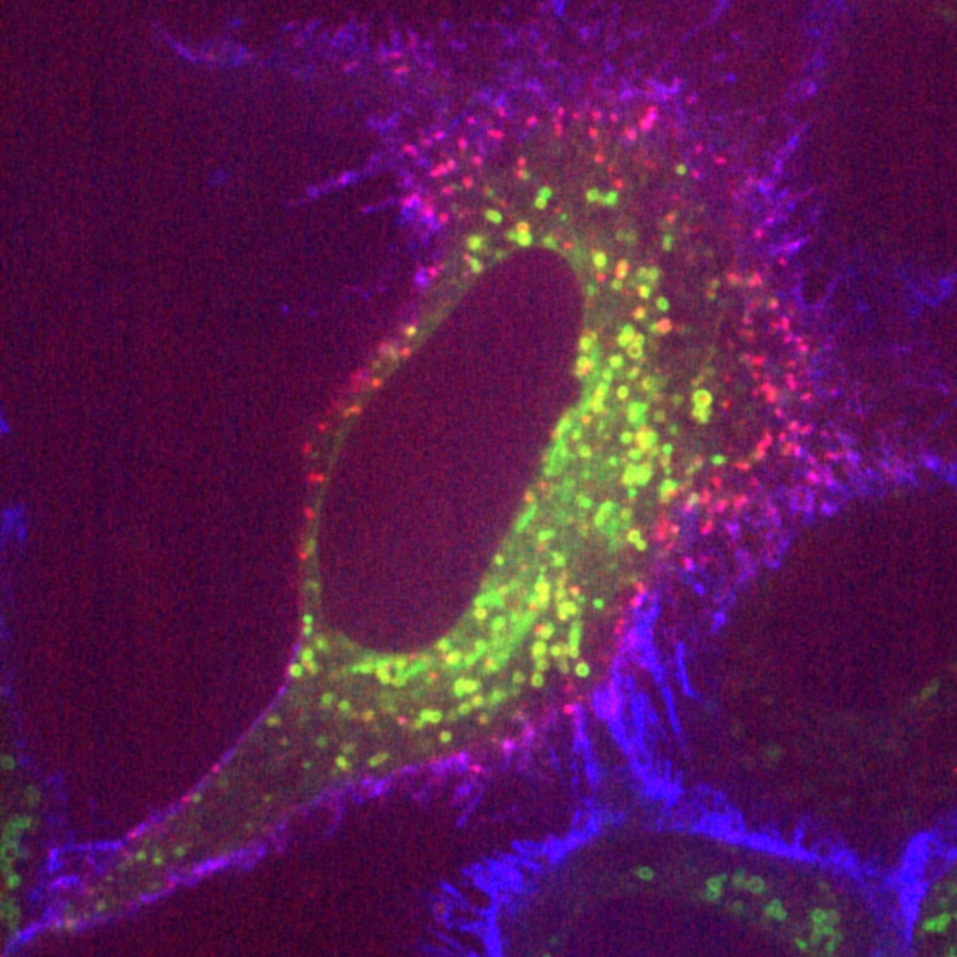
Cells can be thought of as islands – they are closed to the outside world because they have a plasma membrane that doesn’t let anything through. However, cells have evolved tiny portals for things to enter in a highly controlled way. These portals are working all the time. They use a protein called clathrin together with many other proteins in order to work. We are trying to understand how these proteins come together to do this.
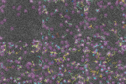
Inside the cell is a busy world with vesicles moving here and there. These vesicles are making sure that important proteins get to the right place at the right time to keep the cell working normally. We have found a new type of vesicle, which is very small (nanoscale). Our lab is trying to figure out how these vesicles are formed and moved around.
Cell division
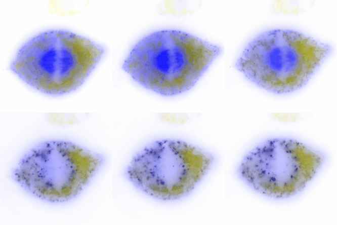
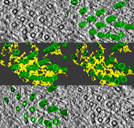
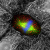
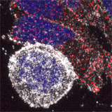
When a cell divides, each of the two new cells must get one copy of the genome each. The cell makes a machine called the mitotic spindle to share the chromosomes (genome) equally between the two cells. This is a very important task: cells that have too few or too many chromosomes can become cancerous. Our lab is trying to figure out how the mitotic spindle does this so efficiently. There are certain fibres within the spindle that pull chromosomes around the cell to share them out. These fibres are made up of many smaller tubules that are held together by “bridges”. We are using powerful microscopes to study these bridges and find out what they are made of.
So far, we have found that some of these bridges are made by four different proteins working together. Three of these proteins are overexpressed in cancer. This means that in patients with these tumours, the cancer cells are making too much of these proteins. We are figuring out how this might cause the tumour, or how it might make the cancer get worse.
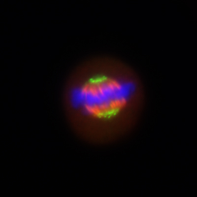
We also working on what happens when cells share the chromosomes incorrectly. Using a microscope that allows us to watch chromosomes inside dividing cells, we can see mistakes in sharing chromosomes. Sometimes, this can lead to micronuclei (very small nuclei) forming in the cell after division, and these are linked to cancer.
